
Review of Innate immunity modulates autoimmunity: type 1 interferon-b
treatment in multiple sclerosis promotes growth and function of
regulatory invariant natural killer T cells through dendritic cell
maturation
Amanda Days
Biomedical Sciences
Introduction to Immunology BIOL 302
Professor Clayton Wright
2/15/2023
- INTRODUCTION
Type 1 interferon- β can be used as therapy for multiple sclerosis due to its nature as an innate cytokine (1). It promotes the use of invariant natural killer (iNKT) cells in the body and the use of this interferon as treatment increases the percent of Vα24+ NKT cells. It should also be noted that the type 1 interferon-β influenced the presenting capabilities of dendritic cells, in turn influencing the activity of the natural killer cells which has been shown to help prevent autoimmune diseases (1).
- HYPOTHESIS
The hypothesis that was put to the test was the idea that type 1 interferon-β could prevent autoimmunity in multiple sclerosis by influencing regulatory mechanisms (1).
- AIMS
To begin the experiment, invariant natural killer T cells were stained with a monoclonal antibody or a combination of the anti-Va24 monoclonal antibody and human CD1d tetramers (1). These cells have important roles in immune regulation. To analyze the dendritic cell phenotype, multiple anti-CD molecules being anti-CD11c, anti-CD80, anti-CD40, and anti-CD1d mAbs were used as well as different devises and instruments that would measure flow and sort the cells (1). DCs were created from monocytes found in the blood while PBMC was incubated in 5% CO2 at 37° C for 2 hours then adherent cells were washed away (1). These cells were then cultured in the presence of human granulocyte-macrophage colony-stimulating factor and rhIL-4 for five days (1).
To expand iNKT cells from peripheral blood mononuclear cells, the total PBMC were cultured in the presence of the invariant natural killer cell antigen, αGalCer, rhIL-7, and rhIL-15. Four weeks after the initial incubation, the iNKT cells were purified using magnetic bead selection to remove the purified iNKT cells from the sample to treat with DCs that were treated with the antigen, αGalCer, for 18 hours then radiated (1). To measure the cytokines and the proliferation of this sample, 1 lCi [3H] thymidine was added per well in the last 16 hours of the culture, which lasted three days (1). The levels of cytokines IFN-γ, IL-10, IL-5, IL-4, and IL-2 were measured by a cytokine kit, BD Cytometric Bead Array, FACSCalibur flow cytometry and a BD CBA Software (1).
- RESULTS
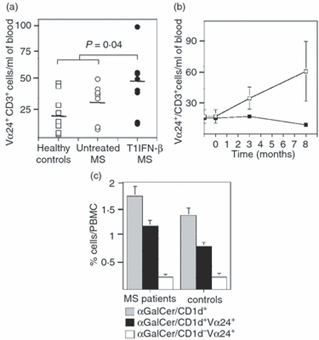
The PBMC had a limited number of iNKT cells, but treatment with T1IFN- β showed a drastic increase in the levels of invariant Vα24+ CD3+ NKT cells in the PBMC versus that of a healthy patient or one before treatment (1). It also had a progressive effect, the number of iNKT cells continuing to grow with time after the initial treatment. Figure 1 shows that the levels of these iNKT cells in patients that were treated had a significant increase (1). It also demonstrates how the levels of these cells increased with time and the most abundant type was the αGalCer/CD1d+ variation (1). It was also confirmed that the treatment itself was needed for the increase in these cells due to the staining and detection of “old” cells after the treatment had discontinued (1).
Figure 1:
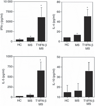
The next major finding was that the treatment encouraged the regulatory iNKT cells to preform their functions. This was seen by the increase in cytokine production of IL-5, IL-4, and IFN-γ after treatment (1). Figure 2 clearly shows the increase levels of the cytokines in the subjects that had undergone treatment. It was also found that the treatment did not directly affect the iNKT cell’s growth and function because the direct combination of the two inhibited the proliferation and secretion of the iNKT cells (1).
Figure 2:
Another finding was the effectiveness of the dendritic cells’ activation of iNKT cells which was more effective in patients that received treatment (1). However, while the DCs had up-regulated expression markers and molecules like CD1d, CD80, and CD40, the cells were not fully mature and downregulated the expression of CD11c as well as failed to secrete cytokines (1).
- DISCUSSION/SUMMARY
This study proved the hypothesis that the treatment of multiple sclerosis with T1IFN- β helps increase the regulatory functions of NKT cells, in turn, improving the condition of the subjects. The treatment increases the DC’s abilities to present antigens to T-cells and secretion of cytokines that aid in antiviral immunity (1). The treatment selectively regulates DC maturation and upregulates costimulatory molecules like CD80 and CD40, as well as CD1d, the iNKT restriction molecule (1). It was also found that the iNKT cells had to balance their functions to have an effective inflammatory immune response as well as prevention of autoimmunity and that T1IFN-β had an effect in improving the function of these iNKT cells (1). Overall, the data proves that the treatment helps improve the growth and functions of iNKT cells which aids in the prevention of autoimmune diseases like multiple sclerosis, which could be expanded to other diseases in the future (1).
Sources:
1) Gigli, G., Caielli, S., Cutuli, D., & Falcone, M. (2007). Innate immunity modulates autoimmunity: Type 1 interferon-β treatment in multiple sclerosis promotes growth and function of regulatory invariant natural killer T cells through dendritic cell maturation. Immunology, 122(3), 409–417. https://doi.org/10.1111/j.1365-2567.2007.02655.x
Leave a Reply