While taking this class, I was also taking Human Anatomy and Physiology 2. Many of the subjects in Cell Biology correlated directly with my anatomy class. For example, when we were discussing cytoplasmic transport in cell biology, I was learning about the endocrine system in my anatomy class. Learning about how hormones are transferred while learning about what hormones are helped me understand the bigger picture and connect all the puzzle pieces, which helped me grow as a student. My particular field of interest is veterinary medicine. Being able to use the knowledge from cell biology and combine it with anatomy and physiology really helped me understand how we work and why our bodies do what they do. While we aren’t exactly the same as animals, the general concept is the same– we are all very complex creatures and there is a lot more to us than most people think!
Scientific Literacy Essay
Inorganic phosphate is a molecule that is critical for cellular metabolism. Structurally, it contains one phosphorus atom and four oxygen atoms and has a negative charge overall. It is found in all living organisms1. When phosphate is not being actively used, it is stored in the skeleton, soft tissues, and blood2. One of inorganic phosphate’s main roles is being a building block for phospholipids and nucleic acids2. Inorganic phosphate is also a crucial part of adenosine triphosphate (ATP), which is a cell’s main form of energy, and plays a role in all forms of cellular metabolism. ATP also plays a large role in signal transduction, which is how a cell communicates.
Cellular metabolism could not work without inorganic phosphates. ATP is made of a molecule called adenine attached to a ribose sugar, which is then attached to a phosphate group3. This molecule can have one, two, or three phosphate groups attached3. Aerobic and anaerobic forms of metabolism use and create ATP. Glycolysis uses four ATP molecules to generate two ATP molecules from glucose3. In lactic acid fermentation, which is an anaerobic form of respiration, two molecules of ATP are generated, as well as in alcohol fermentation, which is also anaerobic3.
This ATP that is created by these processes plays a big role in signal transduction. Signal transduction is a cell’s way of communicating. When a signal is received by a receptor, it sets off a chain of events. The most common pathway for a cellular signal is protein phosphorylation4. A protein detects the signal, called a ligand, and binds it, causing a conformational change and triggering a biochemical cascade4. The protein can then use ATP to create enzymatic action, which eventually leads to cellular changes4. Sometimes, this can even lead to changes in transcription or translation of genes4.
These signals are sent and received across the cellular membrane. Inorganic phosphates make up the main building block of these membranes: phospholipids. Phospholipids are made of a polar head group, consisting of the phosphate group and a choline group or group of similar structure, such as serine, and a nonpolar fatty acid tail, consisting of two fatty acid chains5. This chemical makeup is important for maintaining the integrity of a cell’s structure and helps regulate what goes in and out of the cell5. Multilamellar organelles or bodies are organelles that store and secrete phospholipids6. Once phospholipids are made in the Golgi, they can be transport to a multilamellar body to be saved for later use6. These organelles can be found in multiple cell types, but overall, perform the same function6.
Inorganic phosphates have a wide array of jobs, but these are some of the highest-ranking ones. Without inorganic phosphates, humans would not be able to function like they are able to today. Energy production would not be possible, along with cellular communication and the ability to respond to stimuli. Cells also would not have the ability to maintain membrane integrity due to the phospholipid bilayer surrounding it. Life would be very different without inorganic phosphates.
PXo bodies were recently discovered in a species of fly, Drosophila melanogaster, and have the potential to be considered a new cellular organelle that plays a role in the levels of phosphate in a cell7. In Figure 2, the researchers looked at the biology of the PXo bodies7. They used different fluorescent stains to determine the structures that make up PXo bodies. In panel A, the organelles in question are determined to be round, oval-shaped structures. Panel B helps to further solidify this claim by using immunogold labeling and electron microscopy. The black dots in panels B and C represent where PXo bodies are located and their shape. Panel D shows the number of PXo bodies per square micrometer. The number was highest in the frozen midgut section. In panels E-K, different fluorescent stains are added to help further determine structure. Panel E uses a red marker called LysoTracker Red that detects acidic parts of a cell, usually lysosomes. The green structures are PXo bodies in panels E-K. In the top picture of panel E, there is some overlap (yellow color), which implies PXo bodies are acidic structures. In panel F, red represents Lamp1, which is a lysosome marker. There is no overlap in the first picture of panel F, so PXo bodies are not associated directly with lysosomes. Panel G uses Nile Red as the red marker, which is a lipid dye. There is a significant amount of overlap in the first picture of panel G, implying PXo bodies are associated with lipids. Man II is the red marker used in panel H; it associates with the Golgi. There is no overlap in the first picture, implying there is no association between the Golgi and PXo bodies. In panel I, ConA is used, which is a glycosylation probe. From the overlap in the first picture in panel I, it is inferred PXo molecules are glycosylated. Panel J uses P-Cho, which is a phospholipid tracer. There is significant overlap in the first picture of the panel, which implies PXo bodies are made of phospholipids. Lastly, panel K uses Dextran, which is an endocytosis marker. There is not much overlap in the first picture of the panel, so it is inferred PXo bodies are not involved in endocytosis.
In figure 3, the researchers looked at what effect inhibiting PXo bodies had on inorganic phosphate (Pi) levels in the cytoplasm7. Panel C shows researchers using FRET and FLIPPI to determine if high Pi levels inhibit energy transfer, which it does. In panels D-H, researchers are using heat maps to show when PXo bodies are inhibited (PXo-i in panel E), there are high levels of Pi because FRET is unsuccessful. Inhibiting PXo bodies increased the Pi level in the cytoplasm based on the FRET ratio in panels F and I, so PXo bodies pump Pi out of the cell.
In figure 4, researchers were determining the number and size of PXo bodies7. Panel D shows the diameter of PXo bodies increases when excess Pi is in the cell. When PFA is added and when PXo is inhibited, the diameter of PXo bodies decreased significantly. Panels A-C show the size differences very well. Panels E-G show how the number of PXo bodies decreases when PFA is added and increases when Pi is added. Panel G also shows the size increase of the PXo bodies. Panel J shows the shape of PXo bodies very well; they are oval in shape and numerous in panel G. When PFA is added in panel K, the number and size decrease substantially. Panel H shows this information in graph form; more Pi leads to an increase in number of PXo bodies.
Figure 5 (panels D and E) breaks down the phospholipids in the structure in normal conditions and when PFA is added7. An overall decrease in the number of phospholipids occurs, going from 90.6% to 84.2% phospholipids. Phosphatidylcholine (PC) and phosphatidylethanolamine (PE) are the most prominent in both cases; however, PC decreases slightly when PFA is added. PE, DG, and PA remain the same. PI and TG each decrease by 1% when PFA is added. PS and Cer each increase by 1% when PFA is added. Co disappears when PFA is added, and WE is added, making up 7% of the total structure.
This data shows PXo bodies are unique organelles that have a unique biochemical function. No other organelle has the same exact function as PXo bodies. Their structure is also unique, as they are made up mostly of phospholipids7. This is not surprising since phosphate is used to create phospholipids5. According to a paper written in 2010, these PXo bodies meet the requirements of a cellular organelle- the cell made a cellular membrane for the organelle; the organelle was filled with the correct set of proteins to allow it to do its job; and the cell places the organelle in the correct location and ensure it maintains and segregates the organelle from the rest of the cell8. Overall, the data proves PXo bodies are their own unique organelle with a special function performed by no other organelles.
References
- Pavlov, E., Aschar-Sobbi, R., Campanella, M., Turner, R.J., Gómez-García, M.R., Abramov, A.Y. Inorganic polyphosphate and energy metabolism in mammalian cells. JBC Papers in Press 2010; https://www.jbc.org/article/S0021-9258(19)54998-5/pdf
- Wagner, C.A. The basics of phosphate metabolism. Nephrol Dial Transplant 2024; https://www.ncbi.nlm.nih.gov/pmc/articles/PMC10828206/
- Wikipedia contributors. Adenosine triphosphate 2024; https://en.wikipedia.org/wiki/Adenosine_ triphosphate
- Wikipedia contributors. Signal transduction 2024; https://en.wikipedia.org/wiki/Signal_ transduction
- Bargui, R., Solgadi, A., Prost, B., Chester, M., Ferreiro, A., Piquereau, J., Moulin, M. Phospholipids: identification and implication in muscle pathophysiology 2021; https://www.ncbi.nlm.nih.gov/pmc/articles/PMC8347011/#:~:text=Phospholipids%20%28PLs%29%20are%20amphiphilic%20molecules%20that%20were%20essential,cell%20signaling%2C%20cell%20metabolism%2C%20and%20even%20cell%20pathophysiology.
- Hariri, M., Millane, G., Guimond, M., Guay, G., Dennis, J.W., Nabi, I.R. Biogenesis of multilamellar bodies via autophagy 1999; https://www.molbiolcell.org/doi/pdf/10.1091/mbc.11.1.255
- Xu, C., Xu, J., Tang, H.W., Ericsson, M., Weng, J.H., DiRusso, J., Hu, Y., Ma, W., Asara, J.M., and Perrimon, N. A phosphate-sensing organelle regulates phosphate and tissue homeostasis. Nature. 2023; 617, 798-806. 10.1038/s41586-023-06039-y.
- Murat, D., Byrne, M., and Komeili, A. Cell biology of prokaryotic organelles 2010; https://cshperspectives.cshlp.org/content/2/10/a000422.full.pdf
Portfolio Assignment 3
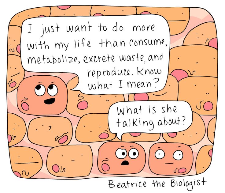
This meme accurately describes how I am feeling at this point in the semester…especially the “what is she talking about” part! It also gives a really brief overview of what we have been learning about this semester—all the jobs of cells!
Portfolio
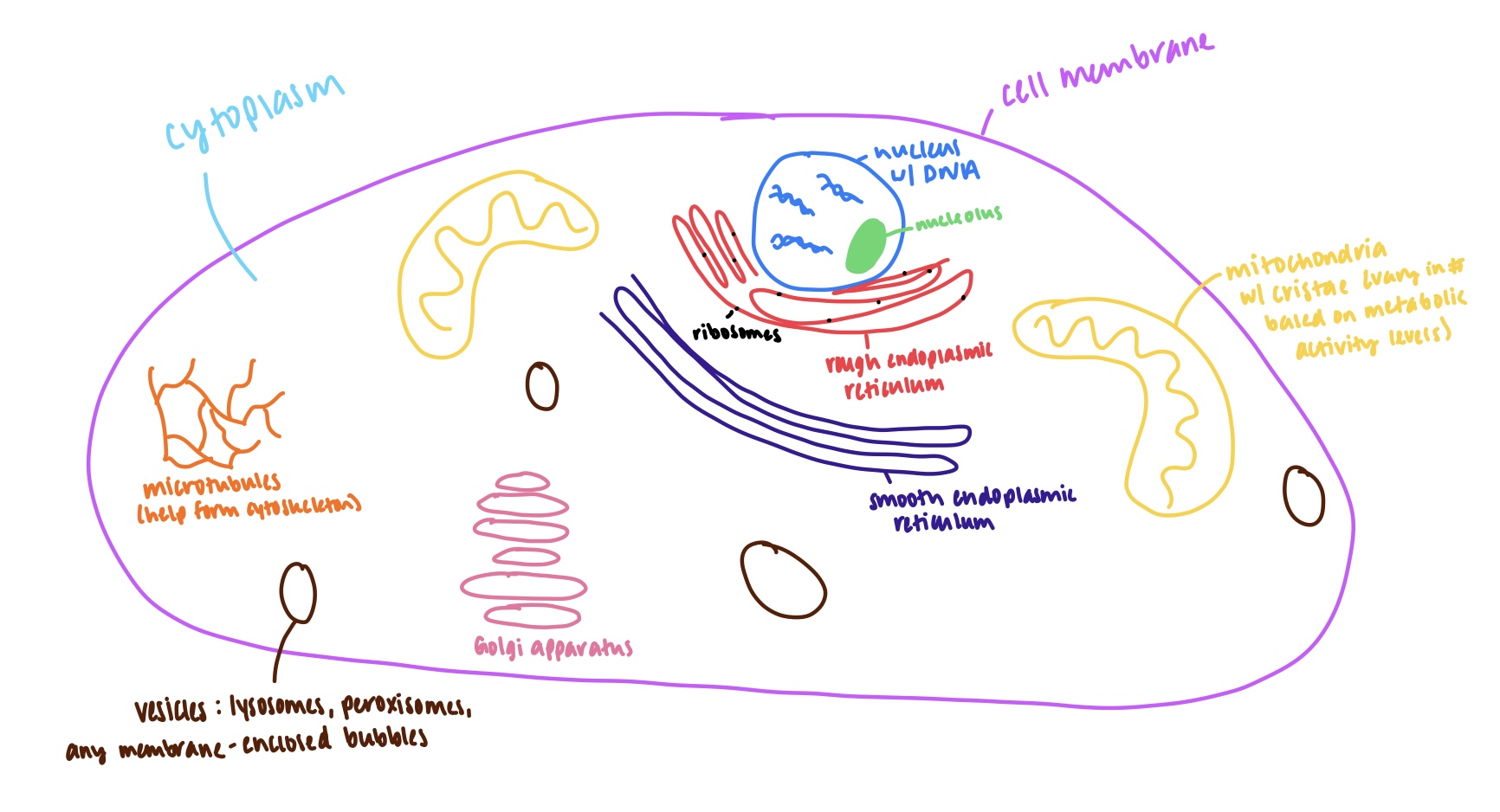
This is my drawing of an animal cell! Although crude, it gives the basic layout of most animal cells. The main structures are labeled for easier identification!
Contact
Please reach out to me at kaseye04@gmail.com with any questions!
Resume
EDUCATION
- Bachelor’s in Animal Science
- Abraham Baldwin Agricultural College
- GPA: 3.88
- Graduated magna cum laude in July 2023
EXPERIENCE
Veterinary Assistant at Animal Health Center
- Collected medical samples for any of the 6 veterinary technicians/assistants
- Administered vaccines and medications
- Held animals for physical examinations for the veterinarian on staff
- Assisted in procedures including surgeries
- Communicated with clients regarding medications, animal care, concerns, or pricing
10 Week Internship at Piedmont Equine Hospital
- Assisted with surgeries and procedures for any of the 8 veterinarians on staff
- Restrained horses for examinations, procedures, and diagnostic imaging
- Collected blood work and ran appropriate lab work
- Administered vaccines and medications
- Helped any of the 11 technicians/assistants with pharmacy and vet truck inventory daily
- Lunged horses for lameness evaluations
Veterinary Intern at Veterinary Care Center
- Assisted 1 large animal veterinarian with procedures and physical examinations
- Restrained horses for examinations
- Administered vaccines and medications
- Drew blood for lab work
Veterinary Assistant at Baytree Animal Hospital
- Collected medical samples for any of the 6 veterinary technicians/assistants
- Administered vaccines and medications
- Held animals for physical examinations for any of the 4 veterinarians on staff
- Assisted in procedures including surgeries
Veterinary Receptionist at Animal Health Center
- Communicated with clients via phone and email
- Scheduled appointments and surgical procedures
- Collected payments
- Answered any questions clients had regarding medications, procedures, or pricing
- Worked with a staff of 1 veterinarian and 6 technicians and receptionists to ensure the clinic ran smoothly
VOLUNTEER EXPERIENCE/LEADERSHIP
Senior Volunteer at Jacobs’ Ladder Therapeutic Riding Center
- Caught, tacked, and groomed 14-18 horses
- Administered medications
- Fed horses and exercised them when necessary
- Coached 12-18 special needs children and adults in horseback riding lessons per week
- Taught between 2-10 new volunteers per day the necessary skills to be successful
- Oversaw between 4-12 volunteers and ensured they were successful
Volunteer Leader for Valdosta First Methodist Church’s Youth and Children’s Ministries
- Taught Sunday morning and Sunday night lessons to anywhere between 5-25 children
- Led a group of approximately 30 youth on several mission trips